Goniometry
The goniometer employs the same light sources as the multispectral microscope, but has an extended capability to measure the scattering properties of a sample by determining the preferred angles light choses to scatter into. The goniometer is therefore important when characterizing the optical properties of human tissue, which in turn lays a foundation for proper interpretation of signals generated by other methods, such as photoacoustic imaging.
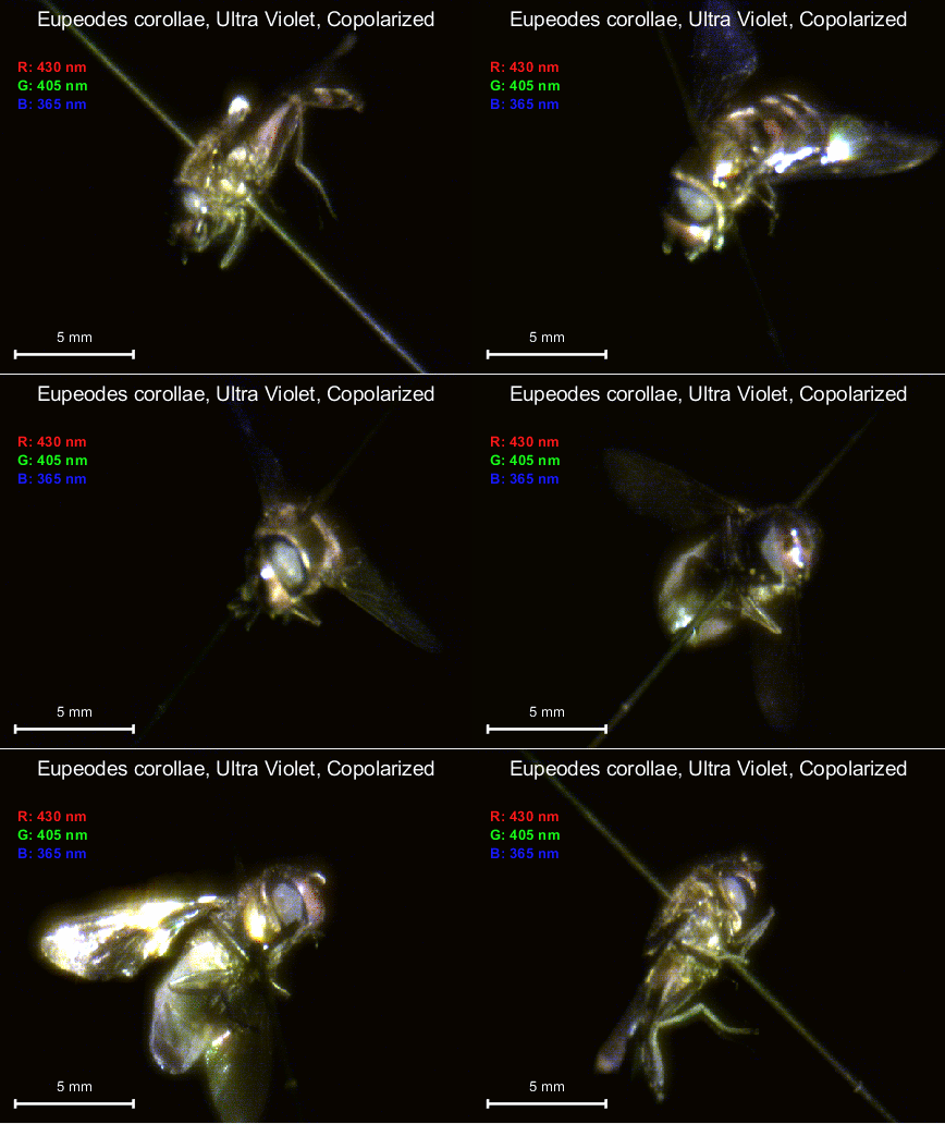
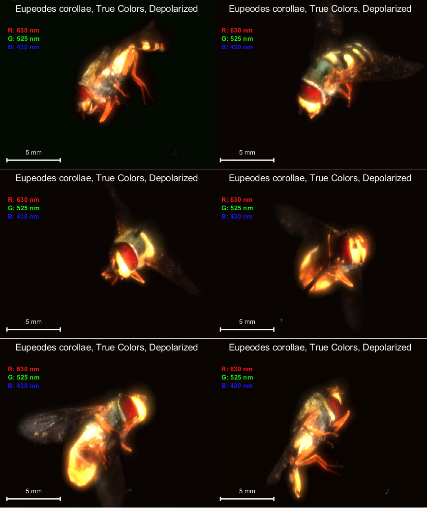
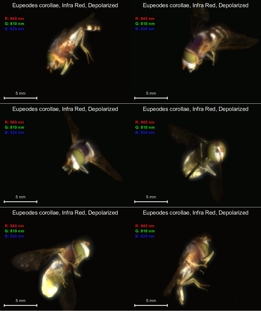
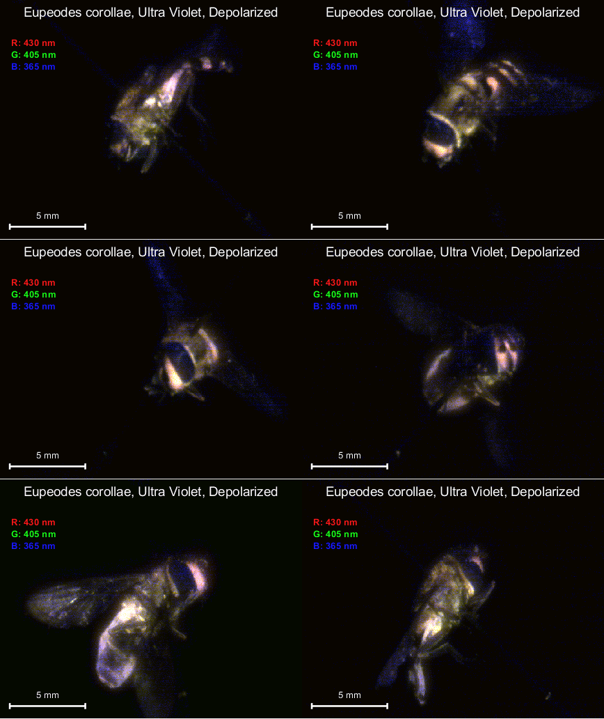
The four images below are from a pilot study and represent the same house-fly taken eith different excitation wavelengths to demonstrate different information. The size of the fly is representative of typical tissue samples that are investigated.


