Breast Cancer
The purpose of this project is to develop and evaluate three non-invasive examination methods for the diagnosis of breast tumors, namely photoacoustics, diffuse reflectance spectroscopy and ultrasound-based tissue characteristics. It is our hope that the methods will be used for primary diagnostic of breast cancer, planning of breast cancer surgery but also to evaluate the given preoperative cancer treatment.
Breast cancer is the most common tumor form among women in Sweden and accounts for about 30% of all cancer cases. The etiology is multifaceted and only in Sweden about 8900 new breast cancer cases are reported every year. Almost half of all cases in Sweden are discovered in connection to the breast cancer screening program where women aged 40-74 years are offered regular mammography. Other cases might be noticed by the patient or the doctor as e.g. a palpable lump due to other symptoms. Triple diagnostics is performed for all suspected breast changes and this includes:
(i) clinical examination;
ii) imaging examination;
iii) cell or tissue sampling, usually with a core needle biopsy (a so-called invasive examination).
In terms of imaging diagnostics (ii), mammography, magnetic resonance imaging (MRI) and ultrasound are effective modalities, but they have their limitations. For example, mammography only provides a structural / anatomical image and the breast is exposed to ionizing radiation. MRI certainly provides dynamic and functional information (in addition to the anatomical image), but it is characterized by its high cost and long examination time. In the case of invasive procedures (iii) there is always the risk of minor bleeding or, in the worst case, infection. A non-invasive method that can provide equal information with a biopsy would mean avoiding these risks.
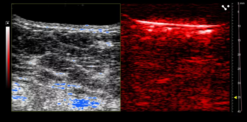
It is of high importance to develop and evaluate non-invasive methods that can provide a traditional anatomical and functional image in a manageable and time-efficient manner, but also information about the molecular composition of the tissue. In this project we will evaluate three non-invasive optical methods that can contribute to improved breast cancer diagnostics, namely photoacoustics (PAI - photoacoustic imaging), diffuse reflectance spectroscopy (DRS) and ultrasound-based tissue characteristics (CFS).
Ultrasound, DRS and PAI differ from other imaging modalities in that the techniques do not use radiation (like X-ray, CT and mammography), magnetic field (MRI) or radioactive isotopes (SPECT / PET). DRS will primarily be used to supplement the photoacoustic image as it provides a more detailed and differentiated measurement of the tissue's optical characteristics in the form of numerical measurement values.
The following specific objectives will be examined:
1a) Can one distinguish between benign and malignant tissue?
b) What are the differences / similarities?
Our hypothesis is that the functional and molecular information, for example, oxygen saturation of suspected tumor tissue, may be a discriminating factor between benign and malignant tissue.
2a) Can an oxygen saturation value be defined to predict malignancy?
b) What other quantitative factors can be a discriminating factor between benign and malignant tumor tissue?
Project participants