Skin Cancer
This project focus on evaluating and optimizing photoacoustic (PA) imaging as a method to detect and outline skin tumors. We are investigating the PA spectral signature of different tumor cells to enable automatic measurement of the tumor borders, and test the developed method in the clinical setting, ultimately developing PA imaging into a diagnostic tool for skin cancer.
When operating tumors in regions with limited tissue, such as the eyelids, it is crutial to not remove too much healthy tissue. Every millimeter one can save plays a major role in the extent of the subsequent plastic surgical reconstruction. At the same time, one wants to be sure that all tumor tissue is surgically removed to reduce the risk of recurrence and the need for further surgery. Currently theses types of tumors are surgically removed, after which the tumor borders are evaluated by histology. If the tumor borders are not enough, the patient is re-operated.
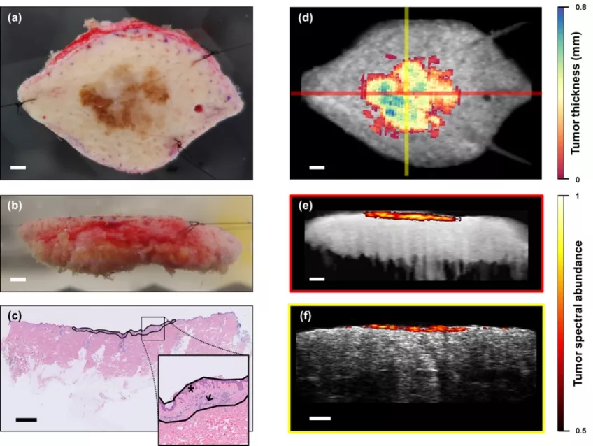
Preoperative, non-invasive delineation of a tumor could improve patient management and eliminate the need for further excision. Diagnosis and tumor border identification with several non-invasive imaging techniques has been attempted, but the results have not been satisfactory. In that context, PA imaging has shown promise: the technique relies on pulsed laser light and optical absorption detected by ultra-high-frequency ultrasound to reveal the molecular composition of tissue at high resolution. However, to date the use of PA imaging has been restricted to pre-clinical experiments and it has not been developed for clinical use. The purpose of the project is to develop PA imaging into a diagnostic tool for skin tumors by identifying the spectral signature of the cancerous cells.
We are currently working on 3 different skin tumor types, melanoma, basal cell carcinoma (BCC) and squamous cell carcinoma (SCC).
Melanoma is one of the most common cancers worldwide and its incidence is increasing. Early diagnosis is crucial for patient survival as, in contrast to metastatic melanoma, early-stage melanoma is highly curable. However, melanoma is difficult to diagnose and stage. Detection is generally based on visual evaluation of lesion morphology, biopsy, and histopathological assessment.
Basal cell carcinoma (BCC) and squamous cell carcinoma (SCC) are the most common forms of non-melanoma skin cancer. BCC rarely spreads to nearby lymph nodes or other parts of the body, but if it is left untreated is can grow into nearby areas and invade the bone or other tissue beneath the skin. SCC on the other hand can like melanoma spread to other parts of the body.
Both melanoma, basal cell carcinoma and squamous cell carcinoma are commonly found in sun-exposed areas, such as the head, neck, and face. Often these areas have limited tissue available for surgery.
Selected publications
Project participants