Machine Learning
Unsupervised skin tumour delineation using hyperspectral imaging and machine learning.
Hyperspectral imaging provides detailed spectral and spatial information of a surface in ways our eyes are not capable of visualizing. We use a custom-made hyperspectral imaging camera from Norsk Elektro Optikk (NEO), capable of providing spectral information between 400-1700 nm. While this allows us to produce images of skin tumours depicting information our eyes normally can’t detect, the data becomes ideal for deep analysis via machine learning.
In this project, we aim to develop a method which enable automatic determination of the tumour borders and further test our developed method in clinical settings. We propose machine learning techniques combined with image segmentation algorithms to tackle these challenges by predicting the actual size of the tumour from hyperspectral images subsequently inferring the optimal size of the tissue that should be removed. We are currently collecting hyperspectral images of skin tumors for training and validating machine learning models that will enable us not only decide easier the size of a tumour but also potentially accurately predict the actual type of tumour.
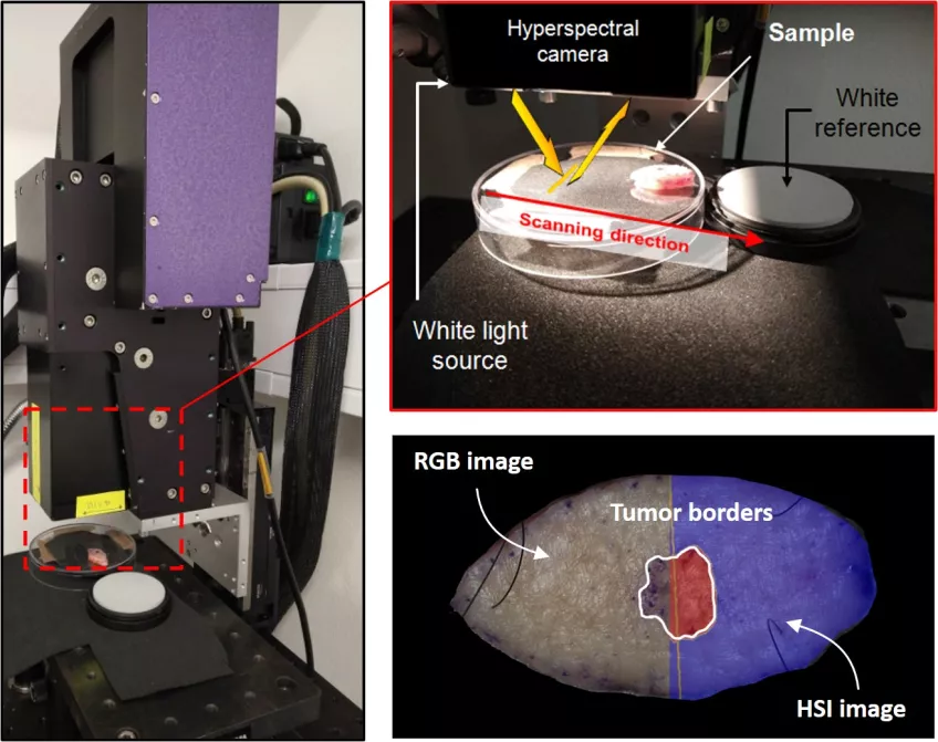
The image below demonstrates the hyperspectral camera in action while capturing hyperspectral images of an excised skin sample with a tumor. Once the data has been imported and subjected to the machine learning techniques under development, the tumor borders found were in strong agreement with that determined from histopathological analysis.
These preliminary results are promising in the quest for unsupervised tumor border delineation with the aid of hyperspectral imaging. The benefits are many, where it could primarily speed up the diagnostic procedure to provide an answer within seconds. Being able to determine tumour borders non-invasively greatly facilitates diagnosis by removing the need for histopathological examination of a biopsy. We estimate that at Skåne Regional Hospital, surgeries could be reduced by 500 a year, should our methods be successfully implemented and used to improve diagnostic accuracy.
This project is a collaboration between the Departments of Ophthalmology and Dermatology at Skåne Regional hospital, and the Department of Theoretical Physics at Lund University.
Project Participants